BEST1 polyclonal, anti-human, mouse, rat
€388.00
In stock
SKU
BS7824
Background:
The retinal pigment epithelium (RPE) and choroid represent a differentiated system of the eye that sustains normal retinal health and function. Best vitelliform macular dystrophy, known as Best disease, is an early-onset autosomal dominant condition in which accumulation of lipofuscin-like material within and beneath the RPE leads to progressive loss of central vision. The lipofuscin-like material in the macular area appears as a yellow mass like the yolk of an egg that later becomes darker and irregular in color, a process known as “scrambling the egg”. Best disease is frequently a reflection of mutations in the Bestrophin gene, which encodes a protein containing four putative transmembrane domains and localizes to the basolateral plasma membrane of RPE cells. Human Bestrophin forms oligomeric chloride channels that are sensitive to intracellular calcium. Missense mutations at the Bestrophin locus reduces or abolishes Bestrophin protein mediated membrane current. The human Bestrophin gene encodes a 585 amino acid protein.
Alternative Name:
ARB, BEST 1, BEST, Best disease, Best macular dystrophy, BEST1, BEST1_HUMAN, Best1V1Delta2, Bestrophin 1, Bestrophin-1, Bestrophin1, BMD, mBest1, RP50, TU15B, Vitelliform macular dystrophy 2, Vitelliform macular dystrophy, Vitelliform macular dystrophy protein 2, VMD 2, VMD2,
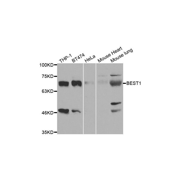
Application Dilution: WB: 1:500~1:2000, IHC: 1:50~1:200
Specificity: BEST1 polyclonal antibody detects endogenous levels of BEST1 protein.
Immunogen:
Recombinant full length Human BEST1.
MW: ~ 68 kDa
Swis Prot.: O76090
Purification & Purity:
The antibody was affinity-purified from rabbit antiserum by affinity-chromatography using epitope-specific immunogen and the purity is > 95% (by SDS-PAGE).
Format:
1mg/ml in PBS with 0.1% Sodium Azide, 50% Glycerol.
Storage:
Store at 4°C short term. Aliquot and store at -20°C long term. Avoid freeze-thaw cycles.
For research use only, not for use in diagnostic procedure.
The retinal pigment epithelium (RPE) and choroid represent a differentiated system of the eye that sustains normal retinal health and function. Best vitelliform macular dystrophy, known as Best disease, is an early-onset autosomal dominant condition in which accumulation of lipofuscin-like material within and beneath the RPE leads to progressive loss of central vision. The lipofuscin-like material in the macular area appears as a yellow mass like the yolk of an egg that later becomes darker and irregular in color, a process known as “scrambling the egg”. Best disease is frequently a reflection of mutations in the Bestrophin gene, which encodes a protein containing four putative transmembrane domains and localizes to the basolateral plasma membrane of RPE cells. Human Bestrophin forms oligomeric chloride channels that are sensitive to intracellular calcium. Missense mutations at the Bestrophin locus reduces or abolishes Bestrophin protein mediated membrane current. The human Bestrophin gene encodes a 585 amino acid protein.
Alternative Name:
ARB, BEST 1, BEST, Best disease, Best macular dystrophy, BEST1, BEST1_HUMAN, Best1V1Delta2, Bestrophin 1, Bestrophin-1, Bestrophin1, BMD, mBest1, RP50, TU15B, Vitelliform macular dystrophy 2, Vitelliform macular dystrophy, Vitelliform macular dystrophy protein 2, VMD 2, VMD2,
Application Dilution: WB: 1:500~1:2000, IHC: 1:50~1:200
Specificity: BEST1 polyclonal antibody detects endogenous levels of BEST1 protein.
Immunogen:
Recombinant full length Human BEST1.
MW: ~ 68 kDa
Swis Prot.: O76090
Purification & Purity:
The antibody was affinity-purified from rabbit antiserum by affinity-chromatography using epitope-specific immunogen and the purity is > 95% (by SDS-PAGE).
Format:
1mg/ml in PBS with 0.1% Sodium Azide, 50% Glycerol.
Storage:
Store at 4°C short term. Aliquot and store at -20°C long term. Avoid freeze-thaw cycles.
For research use only, not for use in diagnostic procedure.
| Is Featured? | No |
|---|
Write Your Own Review