CD55 (Extracellular region) (clone M033), anti-human
€420.00
In stock
SKU
ECM-CM0331
Catalog Number: ECM-CM0331
Size: 100 μl
Isotype: mouse IgG1
Applications: WB, E, ICC, IP
Reactivity: human
Datasheet
Questions? Contact us!
Size: 100 μl
Isotype: mouse IgG1
Applications: WB, E, ICC, IP
Reactivity: human
Datasheet
Questions? Contact us!
Background:
CD55, also known as Decay-Accelerating Factor (DAF) is an inhibitor of the complement system, and is broadly expressed in malignant tumors. In cancer, CD55 has been implicated in tumorigenesis, neoangiogenesis, and metastasis. CD55 may decrease complement mediated tumor cell lysis, inhibit tumor apoptosis, and promote invasive cancer cell motility. These roles in cancer may involve binding to the seven-span transmembrane receptor CD97. In neuroblastoma cells, CD55 contributes to growth of colonies and to invasion of cells, but not to stemness. In neuroblastoma cells, CD55 is upregulated in a small population of cells that are HIF-2α positive. This CD55-positive subpopulation is highly invasive and has low adhesion to fibronectin and collagen. In addition, CD55 expression correlates with poor prognosis in neuroblastoma patients.
Background References
Mikesch JH et al.(2006) Cell Oncol. 28(5-6):223.
Cimmino F et al. (2016) Oncogenesis 5:e212.
Immunogen: Clone M033 was generated from a proprietary antigen related to the extracellular region of human CD55 from the MDA-MB-231 breast cancer cell line.
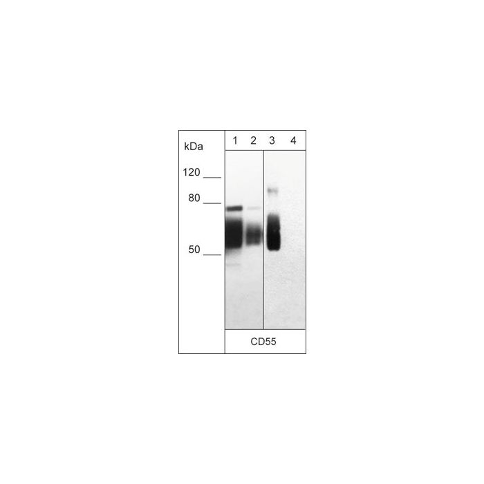
Specificity: Clone M033 was purified using Protein G chromatography. The antibody detects 75-100 kDa* bands corresponding to the molecular mass of CD55 on SDS-PAGE immunoblots of "native" A431, HeLa, MCF7, and MDA-MB-231 cell lysates, but does not detect the denatured form of CD55. The antibody also detects a "native" human recombinant CD55 protein that includes the extracellular region. The antibody can be used for native western blot, immunoprecipitation, protein ELISA, and immunocytochemistry, as well for detecting CD55 in live, unfixed cells.
Application dilution:
ELISA: 1:2000
ICC: 1:100
IP: 1:50
WB: 1:1000
End user should determine optimal dilution for their particular applications and experiments.Western blot membranes were incubated with diluted antibody in 5% non-fat milk, PBS, 0.04% Tween20 for 1 hour at room temperature.
Buffer/Storage:
Mouse monoclonal antibody purified with protein G chromatography is supplied in 100µl phosphate-buffered saline, 50% glycerol, 1 mg/ml BSA, and 0.05% sodium azide. Store at –20°C. Stable for 1 year.
| Is Featured? | No |
|---|
Write Your Own Review

