µCount3D - Fungal Spore and Yeast Counter
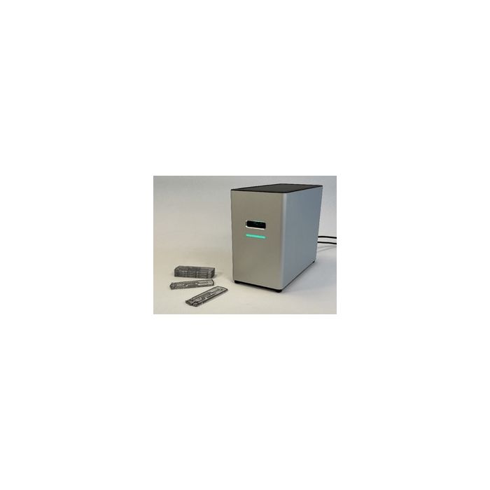
Introducing the µCount3D imaging technology: The µCount3D is developed to count pure cultures of bacteria, fungal spores, and yeasts in liquid samples. µCount3D integrates digital imaging and machine learning to provide precise and reproducible results, using our proprietary FluidScope scanning technology. The FluidScope technology employs a patented and tilted inverted camera technology.
X
You are enquiring about µCount3D - Fungal Spore and Yeast Counter
Automated fungal Spore Counting
Counting fungal spores is an essential task in various scientific disciplines, including mycology, ecology, crop science, food science and medicine. Traditionally, fungal spores have been counted manually under a microscope, but with the advancement of the µCount3D technology, a new, easy and automated counting technology is available.
The µCount3D from BioSense Solutions is developed specifically for microbiology and will accommodate counts of even the smallest of fungal spores.
How we image
The patented FluidScope technology is a tilted camera technology. When images are taken, we get to image a volume instead of a plane. Every image has a height of 150µm and since images overlap we get to create both a vertical and horizontal z-stack. All objects present in this volume, is captured in focus.
For fungal spore counting we image the bottom of a µCassetteF. An image consisting of 400 overlapping images is created and specific deep learning algorithms will count the number of spores present. The µCassetteF sample chambers have an inner height of 200µm allowing fungal spores to settle in just 4 minutes.
The µCassetteF is developed with triplicate sample chambers and the Count3D software will provide fungal spores/ml and supply images for documentation.
µCount3D Specifications
Species: Bacteria, Fungi, Yeast and Algae
Size: H: 20cm W: 10cm D: 20cm
Weight: 3kg
Power consumption: Standby 7W, Running 16,8W
Microbe size: from 0,5µm
Countable Range: 1 x 10⁴ – 1 x 10⁷
3 chamber time to result: ~8 minutes
Output: Organisms/ml, images for documentation, PDF Report
Sample containers: BioSense Solutions triplicate µCassetteF & µCassetteB
Create your own specialized algorithms
Algorithms
In the software you can choose between a range of species. These are all specific algorithms, trained on +2000 sub images from each species. Algorithms are trained to find spores and not mycelium or other particles. Algorithms cover most morphological shapes of fungal spores and if your species is not on the list another algorithm will probably do. Example, if your spore is characterized by having a pill shape, then the Metarhizium algorithm will most likely do a very good job. Many users have a specific background, being product from an antifungal, medium residue, or it can be an environmental sample. In such cases, we always recommend to train a specific algorithm for your specific sample matrix.
Algorithm Development
We work closely with the fungal research community and will be adding new deep learning algorithms for new species and backgrounds continuously. However, we will never cover all needs and you can create your own algorithm using our Annotation Tool. The Annotation Tool It is a stand alone programme, where you annotate the spores in single images. Some can be easy while others will be more complex. Spore populations like Fusarium can exhibit a high degree of heterogeneity and these will take time. Being thorough and consistent is key when annotating. Process is simple – either you annotate or we annotate based on your supplied images. Algorithms developed can further be proprietary or for the common good. On the right is a screen shot from the Annotation Tool training a new algorithm for Cladosporium allicinum in a complex matrix.
QC - Coniothyrium. Counting Chamber Vs. µCount3D
A 2 fold dilution series of Coniothyrium spores was prepared in Eppendorf tubes (0.9% NaCl buffer). First, dilutions was manually counted using a C-Chip, Neubacher Improved, DHC-N01 counting chamber. C-chip was counted using a Zeiss Axioskop 2 plus. One well per dilution. Once counted manually, same samples were pipetted into a triplicate chamber µCassetteF and counted using the µCount3D. Coniothyrium spores counting algorithm was chosen.
Results: Counting results from the manual count was higher than the counts using the µCount3D. There is a good correlation between the 2 dilutions in both counts.
Operator comment: Coniothyrium spores tend to aggregate and it was difficult to count spores in aggregates. The µCount3D Coniothyrium algorithm is trained to count what a human eye can see and if there are aggregates, spores in these are not counted. This probably explains difference between counts.
| Is Featured? | Yes |
|---|
Write Your Own Review