ERK1/2 (clone ), anti-human, mouse
€388.00
In stock
SKU
MB0136
Background:
The activation of signal transduction pathways by growth factors, hormones and neurotransmitters is mediated through two closely related MAP kinases, p44 and p42, designated extracellular-signal related kinase 1 (ERK 1) and ERK 2, respectively. ERK proteins are regulated by dual phosphorylation at Tyrosine 204 and 187 and Threonine 177 and 160 residues mapping within a characteristic Thr-Glu-Tyr motif. Phosphorylation at both the Threonine 202 and Tyrosine 204 residues of ERK 1 and Threonine 185 and Tyrosine 187 residues of ERK 2 is required for full enzymatic activation. The structural consequences of dual phosphorylation in ERK 2 include active site closure, alignment of key catalytic residues that interact with ATP, and remodeling of the activation loop. In response to activation, MAP kinases phosphorylate downstream components on serine and threonine. Upstream MAP kinase regulators include MAP kinase kinase (MEK), MEK kinase and Raf-1. The ERK family has three additional members: ERK 3, ERK 5 and ERK 6.
Alternative Name:
Mitogen-activated protein kinase 3, MAP kinase 3, MAPK 3, ERT2, Extracellular signal-regulated kinase 1, ERK-1, Insulin-stimulated MAP2 kinase, MAP kinase isoform p44, p44-MAPK, Microtubule-associated protein 2 kinase, p44-ERK1, MAPK3, ERK1, PRKM3, Mitogen-activated protein kinase 1, MAP kinase 1, MAPK 1, ERT1, Extracellular signal-regulated kinase 2, ERK-2, MAP kinase isoform p42, p42-MAPK, Mitogen-activated protein kinase 2, MAP kinase 2, MAPK 2, MAPK1, ERK2, PRKM1, PRKM2
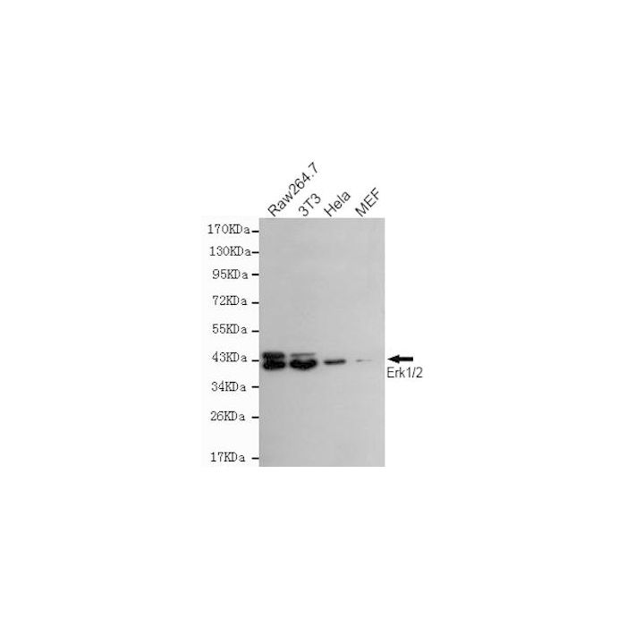
Application Dilution: WB: 1:200~1000
Specificity: This antibody detects endogenous levels of ERK1/2 and does not cross-react with related proteins
Immunogen:
Recombinant full length Human ERK1.
MW: Predicted band size: 42/44KDa, Observed band size: 42/44KDa
Swis Prot.: P27361/P28482
Purification & Purity:
The antibody was affinity-purified from mouse ascites by affinity-chromatography using epitope-specific immunogen and the purity is > 95% (by SDS-PAGE).
Format:
Purified mouse monoclonal in buffer containing 0.1M Tris-Glycine (pH 7.4, 150 mM NaCl) with 0.2% sodium azide, 50%,glycerol
Storage:
Store at 4°C short term. Aliquot and store at -20°C long term. Avoid freeze-thaw cycles.
For research use only, not for use in diagnostic procedure.
The activation of signal transduction pathways by growth factors, hormones and neurotransmitters is mediated through two closely related MAP kinases, p44 and p42, designated extracellular-signal related kinase 1 (ERK 1) and ERK 2, respectively. ERK proteins are regulated by dual phosphorylation at Tyrosine 204 and 187 and Threonine 177 and 160 residues mapping within a characteristic Thr-Glu-Tyr motif. Phosphorylation at both the Threonine 202 and Tyrosine 204 residues of ERK 1 and Threonine 185 and Tyrosine 187 residues of ERK 2 is required for full enzymatic activation. The structural consequences of dual phosphorylation in ERK 2 include active site closure, alignment of key catalytic residues that interact with ATP, and remodeling of the activation loop. In response to activation, MAP kinases phosphorylate downstream components on serine and threonine. Upstream MAP kinase regulators include MAP kinase kinase (MEK), MEK kinase and Raf-1. The ERK family has three additional members: ERK 3, ERK 5 and ERK 6.
Alternative Name:
Mitogen-activated protein kinase 3, MAP kinase 3, MAPK 3, ERT2, Extracellular signal-regulated kinase 1, ERK-1, Insulin-stimulated MAP2 kinase, MAP kinase isoform p44, p44-MAPK, Microtubule-associated protein 2 kinase, p44-ERK1, MAPK3, ERK1, PRKM3, Mitogen-activated protein kinase 1, MAP kinase 1, MAPK 1, ERT1, Extracellular signal-regulated kinase 2, ERK-2, MAP kinase isoform p42, p42-MAPK, Mitogen-activated protein kinase 2, MAP kinase 2, MAPK 2, MAPK1, ERK2, PRKM1, PRKM2
Application Dilution: WB: 1:200~1000
Specificity: This antibody detects endogenous levels of ERK1/2 and does not cross-react with related proteins
Immunogen:
Recombinant full length Human ERK1.
MW: Predicted band size: 42/44KDa, Observed band size: 42/44KDa
Swis Prot.: P27361/P28482
Purification & Purity:
The antibody was affinity-purified from mouse ascites by affinity-chromatography using epitope-specific immunogen and the purity is > 95% (by SDS-PAGE).
Format:
Purified mouse monoclonal in buffer containing 0.1M Tris-Glycine (pH 7.4, 150 mM NaCl) with 0.2% sodium azide, 50%,glycerol
Storage:
Store at 4°C short term. Aliquot and store at -20°C long term. Avoid freeze-thaw cycles.
For research use only, not for use in diagnostic procedure.
| Is Featured? | No |
|---|
Write Your Own Review