FAK (clone ), anti-human, mouse, rat
€428.00
In stock
SKU
BS9850M
Catalog Number: BS9850M
Size: 50 ul, 100 ul
Isotype: rabbit IgG
Applications: WB, ICC/IF, IHC, FC
Datasheet
Size: 50 ul, 100 ul
Isotype: rabbit IgG
Applications: WB, ICC/IF, IHC, FC
Datasheet
Background:
Focal adhesion kinase was initially identified as a major 125 kDa substrate for the intrinsic protein tyrosine kinase activity of Src-encoded pp60. The deduced amino acid sequence of FAK p125 has shown it to be a cytoplasmic protein tyrosine kinase whose sequence and structural organization are unique as compared to other proteins described to date. Localization of p125 by immunofluorescence suggests that it is primarily found in cellular focal adhesions leading to its designation as focal adhesion kinase (FAK). FAK is concentrated at the basal edge of only basal keratinocytes that are actively migrating and rapidly proliferating in repairing burn wounds, and is activated and localized to the focal adhesions of spreading keratinocytes in culture.
Alternative Name:
Focal adhesion kinase 1, FADK 1, Focal adhesion kinase-related nonkinase, FRNK, Protein phosphatase 1 regulatory subunit 71, PPP1R71, Protein-tyrosine kinase 2, p125FAK, pp125FAK, PTK2, FAK, FAK1
Application Dilution: WB: 1:1000-1:2000, ICC/IF: 1:50-1:200, IHC: 1:50-1:200, FC: 1:50-1:100
Specificity: This antibody detects endogenous levels of FAK and does not cross-react with related proteins.
Immunogen:
Recombinant antibody.
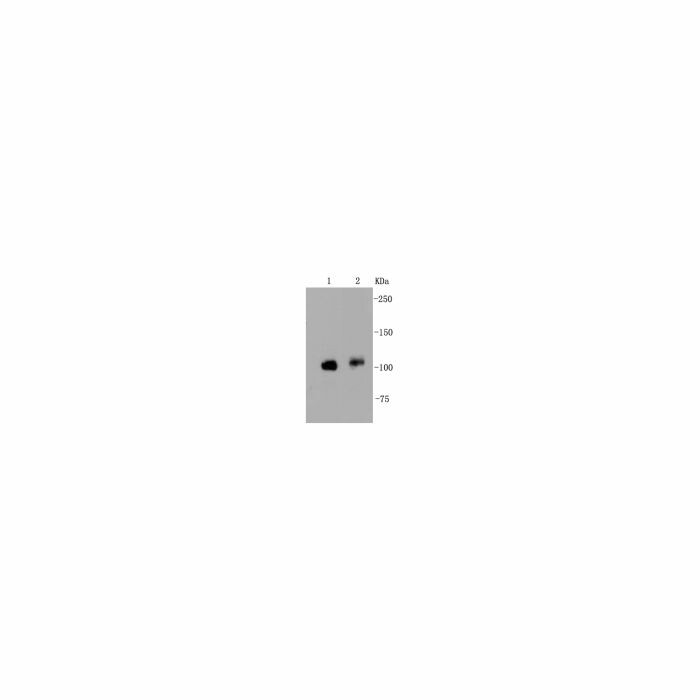
MW: ~ 125 kDa
Swis Prot.: Q05397
Purification & Purity:
Protein A affinity purified
Format:
Recombinant Rabbit Monoclonal Antibody. 1*TBS (pH7.4), 1%BSA, 40%Glycerol. Preservative: 0.05% Sodium Azide.
Storage:
Store at 4°C short term. Aliquot and store at -20°C long term. Avoid freeze-thaw cycles.
For research use only, not for use in diagnostic procedure.
| Is Featured? | No |
|---|
Write Your Own Review

