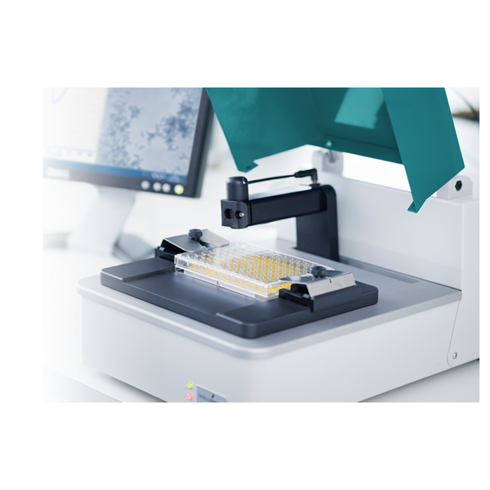
oCelloScope – Automated Microbial Live-Cell Imaging and Analysis
Catalog Number: oCelloScope-1
Quantity: 1 instrument
Product
Request Information - Demonstration
Price on request
oCelloScope – Automated Microbial Live-Cell Imaging and Analysis
Speed
• 250 times more sensitive than a plate reader (OD)
• Measure and visualize down to 5 x 10³ CFU/ml
• Growth kinetic MIC results in few hours Vs 16-20h using BMD
• FluidScope technology scans a full 96-well plate in less than 3 minutes
Early phase morphology
• Compare growth kinetic curves with images from each time-point
• Discover microorganism adaptation strategies
• Capture and quantify morphological changes over time
• Spheroplasts, Filamentation, Co-aggregation, Fungal spore germination
Value
• Full flexibility – use your standard microtiter plates
• You get full software package to use on multiple PC’s
• No expensive annual service contract
• Competitive pricing. Rent or Purchase – just ask for a quotation
Video:
Technology
oCelloScope is a unique live-cell imaging system for sensitive and detailed monitoring of biological growth and development. It’s an easy-to-use automated microscope facilitating high-throughput testing and robust time-lapse studies.
The optical axis is tilted 6.25° relative to the horizontal plane. Such tilting facilitates the scanning of volumes by recording a series of images to form an image stack. The tilting enables more freedom of operation at both high and low concentrations of analyte (microorganisms, mammalian cells, spores, pollen, etc.). The image acquisition process is repeated over time and the time-lapse sequence of best-focus images is used to generate a video.
oCelloScope’s UniExplorer software provides instrument control and data analysis. The optimised algorithms implemented in UniExplorer allow: on-line monitoring of growth and growth inhibition; segmentation of single cells and quantification of morphological features; and antimicrobial susceptibility testing. As oCelloScope enables the visualisation of low analyte concentrations, it also facilitates capturing the early stages in microbial growth that could not otherwise be detected with conventional OD measurements.
How oCelloScope works
For a bacterial suspension in a microwell (Fig. 1), the oCelloScope acquires a sequence of 6.25°-tilted images along the horizontal plane to form an image Z-stack gennerated by the build in algorithms. The Z-stack contains the best-focus image, as well as the adjacent out-of-focus images (which contain progressively more out-of-focus cells, the further the distance from the best-focus position). The acquisition process is repeated over time, so that the time-lapse sequence of best-focus images is used to generate a video.
All the acquired images are saved and used for data analysis. Both the tilted images and the best-focus Z-stack layer can be analysed by the growth kinetic algorithms; the segmentation extracted surface area (SESA) and the two based on measurement of light absorption (i.e., the same principle of optical density, OD, measurements) which are the background corrected absorption (BCA) and the total absorption (TA) algorithms.

• The BCA algorithm provides increased sensitivity and robustness over OD measurements, even at very low or high cell concentrations. To achieve this, the BCA algorithm corrects background intensities with respect to the images acquired at the first time point. This allows obtaining images with an even light distribution, which are used for calculating an intensity threshold. The threshold divides pixels into ‘background’ and ‘objects’. Growth curves are generated based on changes in ‘objects’ so that the effect of background intensities are significantly reduced. The BCA value is calculated as:
BCA value = log10 (Σ(object pixel intensities))
The TA algorithm is designed as an equivalent of OD measurements. During microbial growth, the increasing number of microorganisms will reduce light transmission through the sample and the image will get progressively darker. A darker image is equivalent to a higher TA value. Sensitivity is limited if compared to the BCA algorithm as growth and cell concentration need to be quite considerable before affecting light transmission. The TA value is calculated as:
TA value = log10 (Σ(pixel intensities))
The SESA algorithm uses the simplest method for image segmentation, which is called ‘linear thresholding’. The algorithm identifies all the ‘objects’ in a scan based on their contrast against the background and then calculates the total surface area covered by such objects. Therefore, this method is not based on absorbance but uses the object covered area, so that it is not affected by background intensity changes (such as shadow effects caused by, e.g., condensation on the microtiter plate lid and air bubbles in the culture medium) and can measure microbial growth with high accuracy at very low cell concentrations. However, when more than 20% of the total image area is covered by objects, the SESA algorithm accuracy starts to decline. The SESA algorithm gives faster results compared to conventional OD measurements. The SESA value is calculated as:
SESA value = log10(Σ(object covered area))
Only the best-focus Z-stack layers are analysed by the growth kinetic algorithm based on object-based image analysis (OBIA), which is the segmentation extracted average length (SEAL) algorithm. In general terms, OBIA employs two main processes: segmentation and classification. OBIA groups image pixels into homogeneous ‘objects’, which can have different shapes and intensity scale. The ‘objects’ are also associated with statistics that can be used for classification of such ‘objects’ and include geometry, context and texture.
The SEAL algorithm is specifically designed to detect filamentation of rod shaped bacteria. The SEAL algorithm performs segmentation to identify all the ‘objects’ in a scan and determines their average length. The SEAL algorithm is limited when bacterial cells or filaments are overlapping and may lead to inaccurate determination of bacterial length at high cell concentrations. The SEAL value is calculated as
SEAL value =Σ(object length) / number of objects
The oCelloScope segmentation analysis is also based on OBIA and is performed on the best-focus layer of the Z-stacks to identify and quantify up to twenty different morphological features (e.g., area, eccentricity, symmetry, …) of individual ‘objects’ (or group of objects), such as microbes, spores and cells. Moreover, it is also possible to monitor the development of such morphological features over time with the segmentation kinetics analysis.
For samples that are sedimented to a monolayer at the bottom of the microwell (Fig. 2), all the cells will be in focus along the horizontal plane. Therefore, the image Z-stack will contain the all-in-focus image as well as the adjacent out-of-focus images along the vertical axis. The generation of video and data analysis are the same as shown for samples in suspension.

Schematic showing the oCelloScope optical scanning technology and image analysis applied to a microwell containing bacteria sedimented at the bottom.
| Is Featured? | No |
|---|

