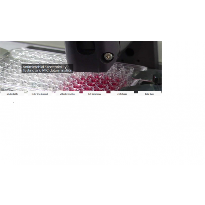
oCelloScope-Bacteria and AMR
Catalog Number: oCelloScope-Bacteria and AMR
Quantity: 1 instrument
Product
Request Information
Price on request
4 times faster than Broth Microdilution (BMD)
The oCelloScope is used by AMR researchers all over the world, including the US CDC to test new or existing drugs against resistant microorganisms.
”All optical screening results were consistent with results from conventional susceptibility tests and were available in 4 h. This decreased the time required to determine antimicrobial susceptibility profiles by 75 to 80%, as conventional testing by BMD requires sufficient visible growth for interpretation of results and has been standardized by CLSI as a 16- to 20-h incubation time for B. anthracis.” McLaughlin, H.P., Gargis, A.S., Michel, P., Sue, D., and Weigel, L.M. (2017). J. Clin. Microbiol. 55, 959–970.
The software interface is intuitive and we typically only need a one hour web intro to get you started. Two modules are primarily used in AMR research – Growth Kinetics and Segmentation. Growth Kinetics is used to screen bacterial or fungal strains for resistance patterns in high throughput. The Segmentation module will take you a step deeper and enable you to analyze and quantify morphological responses.
Join the battle against Antimicrobial resistance with oCelloScope
New resistance mechanisms are emerging and spreading globally, threatening our ability to treat common infectious diseases, resulting in prolonged illness, disability, and death.
Scientists are demanding fast and sensitive methods to discover new antibiotics and push forward our understanding of these resistance mechanisms.
oCelloScope enables scientists to reduce the time-to-result, run faster screening and reduce the manual work-load, by combining growth curves with images and video, through highly sensitive and automated data acquisition and analysis. See movie here.
Case study:
CDC have shown that with oCelloScope you can get 4 times faster time-to-result on Antimicrobial Susceptibility Test for B.anthracis
According to Centers for Disease Control, McLaughlin et al. (2017), oCelloScope can detect the growth of B.anthracis much faster than standard methods, enabling the user to consistently get the data needed for MIC determination within 4 hours, compared to 16-20 hours for standard methods.
This much greater sensitivity of oCelloScope, compared to standard methods, is obtained with the specialized embedded image analysis algorithms, that enables oCelloScope to detect single cells and quantify growth at concentrations down to ~103 CFU/ml.

E. coli growth measured with oCelloScope algorithms, BCA (red) and TA (blue) and Thermo-Fisher Scientific VarioSkan (black). Corresponding images from oCelloScope at time point 0, 120 and 240 minutes.
Accuracy in MIC Determination
Rapid time-to-result is not the only advantage of the oCelloScope.
Fredborg et al., tested 168 antibiotic-bacteria combinations, and with an overall agreement of 96% of the calculated MIC, they concluded that the rapid detection time does not interfere with the validity of the results.
See the protocol for AST and determination of MIC in our application note:
‘Rapid antimicrobial susceptibility testing and determination of MIC value using the oCelloScope’
The UniExplorer software automatically provides the user with a plate overview to quickly evaluate the experiment.
Follow the β-lactam-induced cell morphology changes on Gram-negatives
![]()
oCelloScope combines high sensitivity with real-time imaging, which enables scientist to analyse and follow cell morphology changes.
The special designed SEAL algorithm can detect filamentation of rod-shaped bacteria based on segmentation and extraction of the average bacterial length.
Learn more details and watch the videos in this recent publication from CDC, McLaughlin and Sue (2018), where SEAL was used to measure the average cell length (μm) of B. pseudomallei strains Bp82 and JB039 in the presence and absence of CAZ. 
| Is Featured? | No |
|---|

