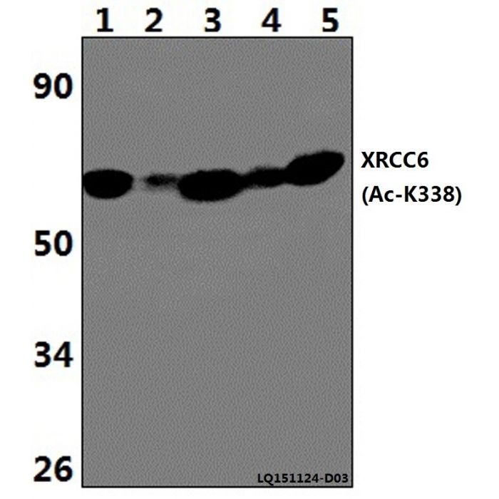
XRCC6 (Acetyl-K338) polyclonal, anti-human, mouse, rat
€388.00
In stock
SKU
BS64099
Background:
The Ku protein islocalized in the nucleus and iscomposed of subunits referred to as Ku-70 (or p70) and Ku-86 or (p86). Ku wasfirst described as an autoantigen to which antibodies were produced in a patient with scleroderma polymyositis overlap syndrome, and waslater found in the sera of patients with other rheumatic diseases. Both subunits of the Ku protein have been cloned, and a number of functions have been proposed for Ku, including cellsignaling, DNA replication and transcriptional activation. Ku is involved in Pol II-directed transcription byvirtue of its DNA binding activity,serving as the regulatory component of the DNA-associated protein kinase that phosphorylates Pol II and transcription factor Sp. Ku proteins also activate transcription from the U1 small nuclear RNA and the human transferrin receptor gene promoters. A Ku-related protein designated the enhancer binding factor (E1BF),composed of two subunits, has been identified as a positive regulator of RNA polymerase I transcription initiation.
Alternative Name:
X-ray repair cross-complementing protein 6, 5'-deoxyribose-5-phosphate lyase Ku70, 5'-dRP lyase Ku70, 70 kDa subunit of Ku antigen, ATP-dependent DNA helicase 2 subunit 1, ATP-dependent DNA helicase II 70 kDa subunit, CTC box-binding factor 75 kDa subunit, CTC75, CTCBF, DNA repair protein XRCC6, Lupus Ku autoantigen protein p70, Ku70, Thyroid-lupus autoantigen, TLAA, X-ray repair complementing defective repair in Chinese hamster cells 6, XRCC6
Application Dilution: WB: 1:500~1:1000
Specificity: XRCC6 (Acetyl-K338) polyclonal antibody detects endogenous levels of XRCC6 protein only when acetylated at Lys338.
Immunogen:
Synthetic Acetylated peptide derived from human XRCC6 around the Acetylation site of lysine K338.
MW: ~ 69 kDa
Swis Prot.: P12956
Purification & Purity:
The antibody was affinity-purified from rabbit antiserum by affinity-chromatography using epitope-specific immunogen and the purity is > 95% (by SDS-PAGE).
Format:
1 mg/ml in Phosphate buffered saline (PBS) with 0.05% sodium azide, approx. pH 7.3.
Storage:
Store at 4°C short term. Aliquot and store at -20°C long term. Avoid freeze-thaw cycles.
For research use only, not for use in diagnostic procedure.
| Is Featured? | No |
|---|
Write Your Own Review

